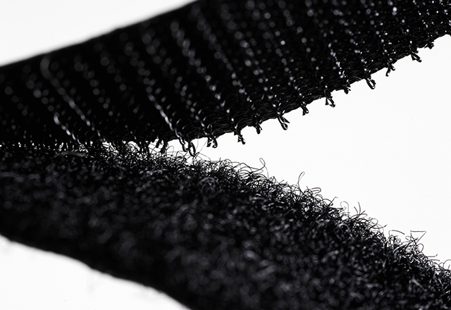
When engineering and biology come together, extraordinary things happen. The meeting of these disciplines has already reshaped our daily lives through biomimicry: the practice of learning from and emulating nature’s time-tested strategies to solve human challenges. Velcro’s tiny nylon hooks, inspired by burdock burrs clinging to a dog’s fur, are standard in clothing, aerospace and medical gear. Japan’s Shinkansen bullet trains feature a nose that is modeled after the kingfisher’s sleek bill and eliminates tunnel “sonic booms,” cutting drag by approximately 30% and lowering energy use. Even the ribbed skin of fast-swimming sharks has guided drag-reducing coatings for ships and hospital surfaces that resist bacterial colonization. Each innovation begins with a simple question — How does nature do that? — and ends with a design that feels inevitable once you’ve seen it.
Yet the deeper we delve into biology, the clearer it becomes that many of medicine’s grand challenges cannot be solved by mimicking form alone. To heal damaged organs, predict drug toxicity or understand whole-body metabolism, we need living systems that faithfully recapitulate human physiology. In this article, I will discuss how the concept of human biomimetics is guiding tissue engineering and the development of microphysiological systems (MPS), but first, let’s confront the problem that drives this work.
The Biomedical Bottleneck
Drug discovery is a marathon of attrition: Roughly 1 of every 10,000 candidate molecules reach FDA approval, after a decade and billions of dollars (1). Much of this attrition comes from the “predictive gap”: Animal models and traditional 2D cell cultures simply can’t replicate the complexity of human tissues, let alone the crosstalk between organs. Therapies that shine in mice often stumble in clinical trials due to unexpected toxicity or lack of efficacy. What we still need is a tenable middle ground: platforms that preserve enough multicellular conversation to reveal the roots of disease while remaining experimentally tractable. Success depends on testbeds that can truly forecast human responses.
Human Biomimetics in Action: Microphysiological Systems
Microphysiological systems answer that call. Often called “organs-on-chips” or “human-on-a-chip,” MPS are fluidic devices that house lab-grown living human cells typically arranged in 3D architectures that simulate dynamic tissue structure and function. In these MPS, fluidic channels perfuse nutrients through controlled flows and impose mechanical cues to mimic the forces those cells experience in the body such as blood-like shear, lung-like stretch, and gut-like peristalsis. Some chips focus on a single organ; others link several organs to recapitulate inter-organ crosstalk. The goal is not to build mini people but to scale biological complexity and capture the principles of communication, feedback and regulation that knit our physiology together.
As a postdoctoral fellow in Dr. Martin Trapecar’s Laboratory of Human Biomimetics at Johns Hopkins All Children’s Hospital in St. Petersburg, Florida, I work where tissue engineering meets biomimetics. Together, as engineers and biologists, we design and 3D print modular devices, embed living scaffolds with primary human cells (including elusive tissue-resident immune populations), and fine-tune flow profiles and mechanics to match each organ’s biomechanics (2). Our combined expertise allows us to cultivate and nurture human tissues that replicate in-body functions, giving us a powerful platform to probe the causal roots of disease and test new therapies with unprecedented precision.
The work that excites me the most within the Laboratory of Human Biomimetics is development of a multiorgan MPS device that imitates systemic glucose regulation. To do this, we have included the major metabolic organs: intestine, pancreas, liver, skeletal muscle, adipose tissue and brain. Collectively, these organs sense circulating glucose, produce insulin, utilize glucose as cellular fuel, and converse through hormones and other signaling molecules. Because metabolic diseases emerge from a web of organ-to-organ signals that no single model can capture, multiorgan MPS offer a human-relevant window into how disturbances in one tissue reverberate throughout other tissues. This insight is indispensable to predict, prevent and personalize treatments for diabetes and other metabolic disorders.
Recreating aspects of human physiology in MPS lets researchers watch disease initiation, progression and treatment with detail once unattainable. Real-time sampling of metabolites flags toxicity early. Live imaging tracks immune-cell patrols as they happen. “Clinical trials on a chip” can test therapies on cells from diverse donors, opening doors to personalized medicine and reducing preclinical timelines. In effect, we are shifting from borrowing nature’s living blueprints to building functional slices of life itself.
Patterning Ourselves After Life: Now a National Priority
Recognizing the predictive gap that hampers drug development, the National Institutes of Health (NIH) has launched a sweeping initiative to prioritize human-based research technologies and reduce animal use (3). A new Office of Research Innovation, Validation, and Application (ORIVA) will steer funding toward organoids, tissue chips, advanced computational models and real-world clinical data, while training study sections to judge proposals on human relevance rather than the tradition of solely using animal models. In short, the NIH is putting its weight behind the same biomimetic platforms that laboratories have been quietly perfecting for years.
This endorsement amplifies a broader shift already under way. Industrial design once forced nature to fit into a linear mindset of straight lines and single-use plastics; biomimicry flipped that script, borrowing the hooks, curves and textured surfaces of life to refine machines. Human biomimetics goes a step further: It builds platforms that behave like us, learn like us and ultimately heal us. The lesson is in humility: Nature is not our competitor but our co-designer, every feather, fin and fungus a prototype refined over eons.
As climate uncertainty, medical complexity and deep-space exploration loom, tomorrow’s breakthroughs may hinge on yesterday’s natural experiments, now accelerated by ORIVA’s mandate. The future will favor those who study not only the stars but also the soil, the skin, the cell. So, when you fasten Velcro or read about a lung-on-a-chip predicting drug safety, remember the quiet revolution unfolding: By reconstructing life’s habitats, cell by cell, perhaps we can finally clear the biomedical bottleneck, replacing long, costly trial and error with miniature ecosystems patterned on life itself.
References:
(1). L. J. Marshall, J. Bailey, M. Cassotta, K. Herrmann, F. Pistollato, Poor Translatability of Biomedical Research Using Animals — A Narrative Review. Alternatives to Laboratory Animals. 51, 102-135 (2023)
(2). Trapecar M. Multiorgan microphysiological systems as tools to interrogate interorgan crosstalk and complex diseases. FEBS Letters. 596(5):681-695 (2022)
(3). National Institutes of Health. NIH to prioritize human-based research technologies. NIH. https://www.nih.gov/news-events/news-releases/nih-prioritize-human-based-research-technologies (2025, April 29).
Related Content
Want to read more from the Johns Hopkins School of Medicine? Subscribe to the Biomedical Odyssey blog and receive new posts directly in your inbox.