
Life is a miracle. But have you ever wondered what the biological trajectory leading to it is? It all begins (partly) in a female’s reproductive organ, ovaries to be scientifically sound. Special cells in the ovaries grow and mature into eggs (oocytes). Each egg has only half the normal number of chromosomes, waiting to combine with the other half from a sperm, culminating in a single-celled zygote, which has a full set of instructions to make a baby. The zygote quickly divides into many smaller cells, forming a tiny ball of cells that attaches to the wall of the mother’s womb (uterus). This is when pregnancy begins.
Now let’s take a step back. “Special cells in the ovaries…. what makes them special?” I asked Dr. Pathak, a postdoctoral researcher in the Spradling Lab at Carnegie Institution for Science -- Department of Embryology. Her eyes lit up and she introduced me to the most fascinating world of developmental biology.
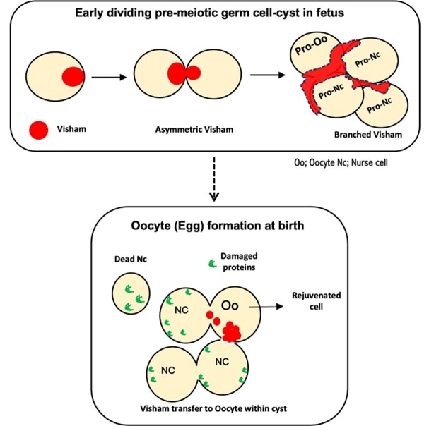
As she explained, inside the ovaries of a female fetus who is awaiting the dawn of life are many tight clusters of equivalent cells, called cysts. These cells are inter-connected — like a group chat where everyone stays linked — but one of them is chosen to be the oocyte while the remaining, named the ‘nurse cells,’ are tasked with nurturing the oocyte.
Over time, the nurse cells transfer their contents to the future oocyte using their inter-cellular connections, whittle down to a fragile remnant, and finally dissolve into nothingness. This is the curse of inequality that is inevitable if life must begin. But isn’t it mysterious as to how this cell-fate is decided, if all the cells were initially equal?
The Spradling lab had been eagerly trying to untangle this question until recently, when Dr. Pathak identified a special structure in mice that she has named the Visham. Visham in Sanskrit means uneven. The Visham, by virtue of its structure, helps organize the cyst so that everything — nutrients, organelles, other cell components — gets funneled into one cell (Pathak & Spradling, 2025).
The story of this unsung and hitherto undiscovered hero doesn’t end here. Now that the oocyte has been marked, it needs to start afresh. “Think of it like rebooting a computer. Before new life can begin, any and all signs of aging or damage in/of the cell need to be completely wiped clean.” explained Dr. Pathak. The Visham helps activate a powerful cellular repair system known as the unfolded protein response, a kind of housekeeping that clears out all damaged proteins and organelles, giving the oocyte a fresh, clean slate.
What left me baffled is that it’s not just in mammals — this biological trick is ancient and well-conserved (Spradling et al., 2022). In insects such as fruit flies, the egg cell clusters build a similar special “communication hub” called the fusome (A., 1901; Huynh & St Johnston, 2004). This hub helps direct important cargo — like energy factories (mitochondria), guiding signals, and other key cell contents — toward the cell destined to become the future egg. It’s nature’s way of making sure every future embryo gets its first care package before the journey even begins.
So next time you ponder how life begins, remember the hidden teamwork and the unsung hero — Visham working behind the scenes, and the selfless sacrifice of numerous nurse-cells that are silently buried under the miracle of life.
References:
A., G. (1901). Origine dell’oocite e delle cellule nutrici nel Dytiscus. Internationale Monatsschrift Fur Anatomie Und Physiologie, 18, 417–484. https://doi.org/10.20848/KONTYU.5.4_145
Huynh, J. R., & St Johnston, D. (2004). The origin of asymmetry: Early polarisation of the Drosophila germline cyst and oocyte. Current Biology, 14(11), R438–R449. https://doi.org/10.1016/j.cub.2004.05.040
Pathak, M., & Spradling, A. C. (2025). Mouse pre-meiotic germline cysts contain fusome-like structure dependent on Dazl that mediates cyst fragmentation and oocyte development. BioRxiv, 2025.07.25.666884. https://doi.org/10.1101/2025.07.25.666884
Spradling, A. C., Niu, W., Yin, Q., Pathak, M., & Maurya, B. (2022). Conservation of oocyte development in germline cysts from Drosophila to mouse. ELife, 11. https://doi.org/10.7554/ELIFE.83230
Related Content
- You Are NOT the Father – How Parthenogenesis Can Explain the Miraculous Pregnancy of a Lone Stingray
- The Powerhouse of the Cell: The Branding of Mitochondria
Want to read more from the Johns Hopkins School of Medicine? Subscribe to the Biomedical Odyssey blog and receive new posts directly in your inbox.