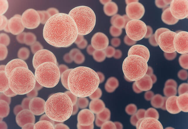
Working with biological samples has its ups and downs. Every Ph.D. student, postdoc and scientific researcher working with cells, tissues, animals or other living organisms knows how tricky it can be. For instance, you might have to work on some weekends or holidays, because these living things usually do not care about the holidays. They cannot say to themselves, “OK, it’s a holiday! Let’s stop growing.” No! They continue to grow and divide. So, you might have to adjust your schedule to suit them. Working with human cells only exacerbates these challenges, as each patient sample behaves in its own unpredictable way. When there is a deadline to meet, I can only pray that these temperamental cultures will grow as expected.
Now, imagine that you are not only supposed to work with living samples from the human body, but you must also grow them in a three-dimensional (3D) culture, called an organoid, instead of in a two-dimensional (2D) culture in a dish. In this case, your challenges could rise exponentially. To start with, on your luckiest days, only 50% of the human tissues would grow and shape themselves as 3D models. Then, when the organoids reach their appropriate sizes, you would have to passage them again and hope the cells do not die. In addition to the cells and tissues themselves, in 3D culturing, you must also struggle with the extracellular matrix (ECM) gels such as collagen and other substrates, which provide support for the organoids. The gels can’t be too liquidy, solid or bubbly. Plus, when you work with collagen, it can’t be too acidic or basic. And these are just a few examples from a long list of do’s and do nots.
But let’s not talk all bad about these little 3D guys! In cancer research, the use of organoids has exploded in recent years, with studies addressing a wide range of research questions in practically every tumor type. If you can grow organoids, then you can have a 3D model of the tissue you are studying that grows from just a small amount of cells (even biopsied tissue), which can come from the original tissue of the human body with all of its characteristics (such as its genetics). This gives researchers the ability to evaluate and study many different features of the tumor cells from a small amount of original tissue while simulating its primary 3D shape. With organoids, researchers can explore tumor cells in a 3D form that could mimic the organization of the cells within the human body. Imagine how much data and information this model of cancer cells can provide compared to just growing cells in 2D. You can explore the cells’ heterogeneity and cell-cell interactions, as well as the invasion of the cells into the surrounding structure. It might be tricky, and you may encounter many challenges. Still, when you overcome all of these challenges and make an organoid, a beautiful circular shape of cancer cells sticking to one another with a defined border, from just a tiny amount of cells, you will be unbelievably excited. Plenty of practice will pay off as you increase your skill, and perhaps even become an expert. After all, science is 90% failure, but the 10% success makes the rest absolutely worth it!
Related Content
- Next Generation Cancer Diagnostics Revolutionize Patient Care
- Foghorn Therapeutics Breaks New Ground in Cancer Epigenetics
- Cancer Diagnosis — It’s in Your Blood
- The Link Between Metabolism and Anti-Tumor Immunity: Implications for Glioma Therapy
Want to read more from the Johns Hopkins School of Medicine? Subscribe to the Biomedical Odyssey blog and receive new posts directly in your inbox.
