γνῶθι σεαυτόν (Know thyself)
Inscribed at the Temple of Apollo at Delphi, this ancient phrase still confronts us thousands of years later. Do we truly know ourselves? What are we? And what is it that makes us us?
In 1637, René Descartes offered a famous answer:
Je pense, donc je suis (I think, therefore I am).
Even if we doubt everything else, Descartes argued, we cannot doubt the existence of the self that doubts and thinks.
Nearly four centuries later, neuroscientist Sebastian Seung proposed a bold hypothesis:
I am my connectome. 1
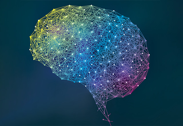
But what is a connectome, and how could it have anything to do with who we are? A connectome is a wiring diagram of the nervous system. Just knowing the properties of individual neurons and how many neurons are in a brain is not enough to understand how the brain works. We also need to know which neurons receive information from which others and where they send their outputs. The brain is a neuronal network, much like the Interstate Highway System: Knowing the properties of individual roads is not enough if we do not know how those roads are connected. A connectome maps these relationships between neurons, and connectomics is the field devoted to reconstructing and analyzing such networks at large scale. Also, understanding these networks may help reveal how neurological and psychiatric disorders disrupt communication across the brain.
Scale matters. In the 1980s, the nervous system of the nematode Caenorhabditis elegans became one of the first to be reconstructed nearly in its entirety, largely because it contains only about 300 neurons.2 Mammalian brains, however, are on a completely different scale. In 2025, the MICrONS project released a reconstruction of roughly 1 cubic millimeter of mouse visual cortex.3 That tiny volume (about the size of a grain of sand) contained more than 200,000 cells and roughly 500 million synapses.
This is one reason scientists turned their attention to the fruit fly. The fruit fly has a nervous system smaller than that of a mouse or a human, yet it walks, flies, feeds, courts, fights, plays, sleeps and learns. In 2024, the FlyWire Consortium published a nearly complete connectome of the adult female fruit fly brain in Nature. The dataset contained approximately 139,000 neurons and 54.5 million chemical synapses.4 Recently, a connectome of the complete central nervous system of an adult male fruit fly was released.5 For the first time, we can begin to trace complete pathways from sensory input, through the brain, all the way to motor output across an entire adult insect nervous system.
And now, people are beginning to place connectomes into computational models and ask whether we can simulate how a nervous system behaves. Researchers have already used the fruit fly connectome database to calculate neural responses to simulated stimulation.6 Other studies have dynamically modeled circuits in the ventral nerve cord, predicted networks capable of generating rhythmic motor activity required for walking, and then tested those predictions experimentally using optogenetics.7 Just look at the countless fruit fly brain simulation memes now circulating online. Virtual fly nervous systems are solving Rubik’s Cubes, playing Minecraft, DJing, scrolling through Reels, YouTube Shorts, and Facebook (just like the rest of us), and even being uploaded into physical machines that move through the real world.
Does this mean that having a connectome is enough to understand how a brain works? Before answering that question, let’s imagine a photograph. A person is standing in the frame. There is mud on the front of his shoes and breadcrumbs on his hands.
If you were Sherlock Holmes, perhaps you might say:
“The mud on his shoes suggests he passed through the nearby construction site. The crumbs on his hands and mouth, together with the stains on his clothes, indicate that he was at William’s Bakery on 120th Street only moments ago. And judging from his expression and the way he is walking, he appears to be desperately looking for a restroom. The only public restroom nearby is in that building. That is where he is going.”
But we are not Sherlock Holmes. And neuroscientists are not Sherlock Holmes either. From a single photograph, we cannot know with certainty where this person came from, why he is here, or where he will go next.
A connectome presents a similar problem. Current connectomes are static snapshots of structure. The wiring diagram of C. elegans has been known for decades. Yet this does not mean that we fully understand every one of its behaviors or all the neural mechanisms that generate them.
An electron-microscopy connectome can show how the nervous system was structurally connected at a particular moment. But neuronal structure alone cannot tell us everything. It does not fully tell us how strong each connection was at that moment, how excitable each neuron was, which neuromodulators were being released, what the animal had just experienced, whether it was hungry or sleepy, or what would change in the next moment. The same wiring diagram can produce very different outcomes depending on the internal state of the system. A living thing, or rather life itself, is not a photograph. It is a movie. For now, obtaining a synapse-level connectome generally requires fixing the brain, cutting it into extremely thin sections, and imaging those sections with electron microscopy. That means we cannot yet continuously observe the changing connectome of the same living brain over time.
One day, we may no longer be limited to a static photograph. There was a time when reconstructing nearly 140,000 neurons and tens of millions of synapses in a fruit fly brain also seemed unrealistic. Yet advances in electron microscopy and AI, together with large-scale human collaboration, made it possible. We may be able to observe something closer to a dynamic connectome, one that captures not only structure but also how that structure changes over time. Also, technologies developed in fruit flies may eventually extend to humans. Further into the future, we might no longer need to reconstruct connectomes by slicing apart a dead brain. Novel scanning technology may allow us to map neural connectivity in a living brain. If that ever becomes possible, each of us could one day have a personal connectome. But even then, the same question would remain. Would that map truly be me? Or would it still be nothing more than an unbelievably detailed trace left behind by the person I happened to be at one particular moment?
Am I my connectome? For now, I would like to leave that question open. Current connectomes still seem to capture only part of what makes us who we are. But my answer might change if we could one day read not just the wiring of a brain, but how that wiring changed over a lifetime.
What are we? Then perhaps we could finally say:
We are our connectomes.
References
- Seung, S. “I Am My Connectome.” TEDGlobal, 2010.
- White, J. G., Southgate, E., Thomson, J. N., & Brenner, S. “The structure of the nervous system of the nematode Caenorhabditis elegans.” Philosophical Transactions of the Royal Society of London. Series B, Biological Sciences 314, 1–340 (1986). doi:10.1098/rstb.1986.0056.
- The MICrONS Consortium. “Functional connectomics spanning multiple areas of mouse visual cortex.” Nature 640, 435–447 (2025). doi:10.1038/s41586-025-08790-w.
- Dorkenwald, S., Matsliah, A., Sterling, A. R., et al. “Neuronal wiring diagram of an adult brain.” Nature 634, 124–138 (2024). doi:10.1038/s41586-024-07558-y.
- Berg, S., Beckett, I. R., Costa, M., et al. “Sexual dimorphism in the complete Drosophila male central nervous system connectome.” Cell 189, 5504–5526.e15 (2026). doi:10.1016/j.cell.2026.08.015.
- Shiu, P. K., Sterne, G. R., Spiller, N., et al. “A Drosophila computational brain model reveals sensorimotor processing.” Nature 634, 210–219 (2024). doi:10.1038/s41586-024-07763-9.
- Pugliese, S. M., Chou, G. M., Abe, E. T. T., et al. “Connectome simulations identify a central pattern generator circuit for fly walking.” bioRxiv (2025). doi:10.1101/2025.09.12.675944.
Related Content
- ‘This Is Your Brain on Drugs’ — Well, on Marijuana at Least
- What Birds in Love Teach Us About How the Brain Processes Competing Motivations
- How the Brain Makes Sense with Mental Maps
Want to read more from the Johns Hopkins School of Medicine? Subscribe to the Biomedical Odyssey blog and receive new posts directly in your inbox.